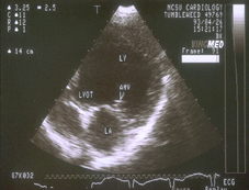
Ultrasonography (also called ultrasound or sonography) is a noninvasive, pain-free procedure that uses sound waves to examine a pet’s internal organs and other structures inside the body. It can be used to evaluate the animal’s heart, kidneys, liver, gallbladder, and bladder; to detect fluid, cysts, tumors, or abscesses; and to confirm pregnancy or monitor an ongoing pregnancy.
We may use this imaging technique in conjunction with radiography (x-rays) and other diagnostic methods to ensure a proper diagnosis. Interpretation of ultrasound images requires great skill on the part of the clinician.
The ultrasonographer applies gel to the surface of the body and then methodically moves a transducer (a small handheld tool) across the skin to record images of the area of interest. The gel helps the transducer slide more easily and create a more accurate visual image.
The transducer emits ultrasonic sound waves, which are directed into the body toward the structures to be examined. The waves create echoes of varying degrees depending on the density of the tissue and amount of fluid present. Those waves create detailed images of the structures, which are shown on a monitor and recorded for evaluation.
Ultrasound does not involve radiation and has no known side effects. For some animals and in certain situation, sedation or anesthesia may be required to allow for a faster and less stressful procedure. The hair in the area to be examined usually needs to be shaved so the ultrasonographer can obtain the best result.
If you have any questions about our ultrasonography service or what to expect during your pet’s procedure, please don’t hesitate to ask.
Our Toronto hospital has an ultrasound unit on premiss allowing for easy and quick bookings of this procedure.
